

In Log Entry #6 I wrote about what the experience was like for me during the actual CCSVI procedure. Here, in Log Entry #7, I’ll get into the details about the abnormalities that were found, and the actions taken to correct them. To catch up on my complete CCSVI Diagnosis and Treatment log, click here (and then scroll down).
What an amazing experience it was.
Right Internal Jugular
An angiogram, which is an X-ray test using a special dye and camera to take pictures of the blood flow in the vein, was performed in the right internal jugular vein. This revealed a short segment stenosis adjacent to the confluence of the brachiocephalic vein. The diameter reduction associated with this stenosis was approximately 60%. The more important number, relative to flow restriction, is the cross sectional area reduction which in this case was approximately 85%.
Multiple collateral veins were noted at this location. Collateral veins indicate attempts by the body to establish bypass flow around an obstruction. Dr. Sclafani inserted the intravascular ultrasound device (IVUS) and confirmed the stenosis. A balloon was inserted, and the vein was dilated multiple times. Another angiogram was performed after dilation which indicated improved flow, diminished flow through collaterals, and no stenosis. Note that the nominal diameter of this blood vessel was 12 mm. Dr. Sclafani prefers to slightly over-dilate the vessel in these instances, and so a 14mm x 4 cm balloon was used. Click on any image to enlarge it.


From left to right, the first image shows the stenosis during angiogram. The second image shows the balloon partially inflated. Note evidence of the stenosis resisting dilation. The third image shows the vein fully dilated.
Left Internal Jugular
An angiogram was performed in the left internal jugular vein. This revealed a stenosis at the confluence with the subclavian vein. The degree of narrowing was similar to that in the right internal jugular vein, approximately 60% diameter reduction and 85% cross sectional area reduction. Collateral veins were noted at this location. The IVUS probe was inserted and confirmed stenosis. A 14 mm x 4 cm balloon was inserted, and the vein was dilated multiple times. Another angiogram was performed after dilation which indicated improved flow, diminished flow through collaterals, and no stenosis.

The above image shows the stenosis during angiogram.


The above images show the balloon during inflation. Note evidence of the stenosis resisting dilation.

Above, angiogram after dilation, showing improved flow through the vein.
Azygos Vein
An angiogram was performed and indicated an abrupt lack of filling approximately 4 cm from the junction between the azygos and the superior vena cava. Dr. Sclafani inserted the IVUS device, which revealed a valve that was failing to completely open. This area was dilated with a 10 mm x 2 cm balloon. Note that the nominal vessel diameter at this location was approximately 8 mm. Another angiogram was performed, and improved flow was noted.

Above is an image of a shorter balloon being used to dilate the azygos at a relatively sharp turn in the vein.
Vertebral Veins
An angiogram was performed on the right vertebral vein. Stenosis was noted, but not treated. This is a smaller vein, and Dr. Sclafani was not comfortable performing angioplasty on it.
An angiogram was performed on the left vertebral vein, indicating that the vein branched into two smaller veins, which is abnormal. No treatment was attempted.
IVUS Images
Dr. Sclafani is innovating the use of IVUS technology in CCSVI diagnosis. I’ll have to take Dr. Sclafani’s word regarding what these images show. Maybe they are like the 3-D pictures that were so popular in the 1990’s. You remember- stare at them long enough and you see dolphins jumping over rainbows (or am I thinking about something else altogether). Anyway, here are some example images.


Above, the image to the left shows narrowing of the left internal jugular vein. The image to the right shows the same vein distended during inspiration (breathing in). Wait! I think I see it now. Do you?


The images above show an incompletely opening valve. I kinda see it.
Significance of findings and treatment
Most of the public discourse regarding CCSVI has been regarding the internal jugular veins and the azygos vein, so I will address these first. All three veins showed some type of stenosis, which is a significant finding. The fact that all three stenoses were repaired after angioplasty is potentially significant for my disease progression.
On the other side of the equation, it is unknown whether the repair of these stenoses will be permanent. Furthermore, since most of my lesion load is in the cervical spine, it is unclear how much benefit I will see from even permanent treatment of stenoses in the internal jugular veins.
The findings of stenosis in the vertebral veins may be significant, given the location of my primary lesion load. In my situation, the vertebral veins may be a more important factor than they are in patients with a lesion load primarily in the brain. This remains to be seen.
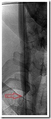
Below is an MRI image of my cervical spine. This is the problem we are trying to solve (or at least halt the further progression of). Arrows indicate MS lesions.

The Bottom Line
I had abnormalities in five out of five veins examined- all are blood vessels that drain my central nervous system. Therefore, it can be said that I had significant CCSVI. The three larger veins were all treated with angioplasty. The two smaller vertebral veins were not treated. One day I hope that we can attempt to treat at least one of these vertebral veins.
Only time will tell if Dr. Sclafani’s efforts were enough to slow or even stop the progression of my MS. I can only hope.
If I could help our re : Chiefumtaga, the internal jugular valve stenosis was at the juncture of the left internal jugular and the subclavian (below the clavicle) artery. The image correlates perfectly.
As well, you may be assuming that the x-ray image is straight on. In fact, in these angio suites the machine rotates at many angles. Because the clavicle is anterior (above) the veins being examined, when the machine is angled it is projected differently than you would see in diagramed anatomy.
It takes a lot of years to learn how to read radiology images, they can be quite deceptive. I was an x-ray tech for 18 years before going off on disability and learned something new every day.
God bless you. Thank you for sharing.
During the procedure were you sedated or given anaesthesia or nothing? I'm curious. (I know that MS persons should stay away from anaesthetics if at all possible.)
Also, I hope your loved ones will help you out with your log…just in case the MS fatigue and your associated symptoms tell you you need a rest.
Hugs,
Thel
Thel,
Thank you for commenting. The interventional radiologists like to have their patients awake for these procedures if at all possible. Throughout the procedure they ask you to do things like breath in, exhale, hold your breath, and push like you are having a bowel movement.
Thank you Anonymous for your post and I wish more people will participate on this boards. I can understand the doctor not doing the procedure but we are not being objective. I have the impression that the comments are scaring the patients and not educating the market. All procedures have a danger factor but what concerns that medicine is taking that privilege from the patients.
The report is excellent with great visuals. A lot of people need to understand CCSVI. A lot of patients say the same that the next feels like nothing.
What is the scope of this procedure under the new Health Reform in the United States?
I had to move back to Puerto Rico because I was diagnose in the United States but MS was a Pre-Existing condition and I could not get an affordable Health Insurance. In Puerto Rico the transportation services are deplorable and in the meantime MS Patients where left stranded in Miami.
In Puerto Rico, I had to wait a year for the Health Care Reform to kick in and now I have to wait until 2014. I got many answers on my condition then but I left on June 2007. Here they make the patient feel like I am lost just like when I left Florida but I got so many answers on my health but here medicine wants you to slave the patient to the condition. Services in the United States with the economy are deplorable too. In a way is moving to a way of living like Puerto Rico but that the decision makers on health issues do not accept reality. Therefore, you have the reality of the patients and the duality of doctors.
Here in Orlando I had to wait 2 years to finally see a Neurologist. The do not want patients with no insurance. I was paying to see him but they do not want to take paying patients.
Eileen,
I don't expect the new health reform to have much impact on CCSVI, one way or the other. In my book, the new bill helps some things, hurts other things, and won't have much effect for years to come. But I must admit, I simply haven't studied it in detail.
I've been fortunate in my lifetime to always have insurance, so I can't comment on the problems that you've had. I certainly hope that the new medical bill will help you…eventually.
Thanks for stopping by and commenting.
You claim not to expect the new health reform to have much impact on CCSVI, one way or the other and if your book says that the new bill helps some things, hurts other things. I imagine that it will not be worse than the Health Reform in Puerto Rico. Thank you for admitting that you have not studied it in detail.
Thank God that you been fortunate to always have insurance. I had Health Reform in Puerto Rico and at least I had Health Care but there the transportation is terrible. I have my hope that the problems that I have will be taken care with the new medical bill eventually but I wonder how long takes for a new procedure to be accepted in the United States.
How long does it takes for the poor society to benefit in the United States from a new procedure?
Mitch,
Did you ever got have anything from Vocational Rehabilitation? Not every patient with MS had the good luck you had.
I have a friend from Chile an Engineering too and he had the procedure after the quake and he is doing great.
There is still a lot we can do. I do to engage as a spokesperson in Puerto Rico. It is great you are blessed with your wife. I notice that the economy will be a challenge to all but more the poor. Even MS patients do not have proper advocating services. You attitude is the same for most MS patients and I have found that learning the law helps a lot. I am currently working on my Bachelor of Science on Paralegal Studies. I am happy that you still Enjoying the Ride of Life.
I wonder why the Neurologist have so much power.
Thank you for reading.