

In Log Entry #6 I wrote about what the experience was like for me during the actual CCSVI procedure. Here, in Log Entry #7, I’ll get into the details about the abnormalities that were found, and the actions taken to correct them. To catch up on my complete CCSVI Diagnosis and Treatment log, click here (and then scroll down).
What an amazing experience it was.
Right Internal Jugular
An angiogram, which is an X-ray test using a special dye and camera to take pictures of the blood flow in the vein, was performed in the right internal jugular vein. This revealed a short segment stenosis adjacent to the confluence of the brachiocephalic vein. The diameter reduction associated with this stenosis was approximately 60%. The more important number, relative to flow restriction, is the cross sectional area reduction which in this case was approximately 85%.
Multiple collateral veins were noted at this location. Collateral veins indicate attempts by the body to establish bypass flow around an obstruction. Dr. Sclafani inserted the intravascular ultrasound device (IVUS) and confirmed the stenosis. A balloon was inserted, and the vein was dilated multiple times. Another angiogram was performed after dilation which indicated improved flow, diminished flow through collaterals, and no stenosis. Note that the nominal diameter of this blood vessel was 12 mm. Dr. Sclafani prefers to slightly over-dilate the vessel in these instances, and so a 14mm x 4 cm balloon was used. Click on any image to enlarge it.


From left to right, the first image shows the stenosis during angiogram. The second image shows the balloon partially inflated. Note evidence of the stenosis resisting dilation. The third image shows the vein fully dilated.
Left Internal Jugular
An angiogram was performed in the left internal jugular vein. This revealed a stenosis at the confluence with the subclavian vein. The degree of narrowing was similar to that in the right internal jugular vein, approximately 60% diameter reduction and 85% cross sectional area reduction. Collateral veins were noted at this location. The IVUS probe was inserted and confirmed stenosis. A 14 mm x 4 cm balloon was inserted, and the vein was dilated multiple times. Another angiogram was performed after dilation which indicated improved flow, diminished flow through collaterals, and no stenosis.

The above image shows the stenosis during angiogram.


The above images show the balloon during inflation. Note evidence of the stenosis resisting dilation.

Above, angiogram after dilation, showing improved flow through the vein.
Azygos Vein
An angiogram was performed and indicated an abrupt lack of filling approximately 4 cm from the junction between the azygos and the superior vena cava. Dr. Sclafani inserted the IVUS device, which revealed a valve that was failing to completely open. This area was dilated with a 10 mm x 2 cm balloon. Note that the nominal vessel diameter at this location was approximately 8 mm. Another angiogram was performed, and improved flow was noted.

Above is an image of a shorter balloon being used to dilate the azygos at a relatively sharp turn in the vein.
Vertebral Veins
An angiogram was performed on the right vertebral vein. Stenosis was noted, but not treated. This is a smaller vein, and Dr. Sclafani was not comfortable performing angioplasty on it.
An angiogram was performed on the left vertebral vein, indicating that the vein branched into two smaller veins, which is abnormal. No treatment was attempted.
IVUS Images
Dr. Sclafani is innovating the use of IVUS technology in CCSVI diagnosis. I’ll have to take Dr. Sclafani’s word regarding what these images show. Maybe they are like the 3-D pictures that were so popular in the 1990’s. You remember- stare at them long enough and you see dolphins jumping over rainbows (or am I thinking about something else altogether). Anyway, here are some example images.


Above, the image to the left shows narrowing of the left internal jugular vein. The image to the right shows the same vein distended during inspiration (breathing in). Wait! I think I see it now. Do you?


The images above show an incompletely opening valve. I kinda see it.
Significance of findings and treatment
Most of the public discourse regarding CCSVI has been regarding the internal jugular veins and the azygos vein, so I will address these first. All three veins showed some type of stenosis, which is a significant finding. The fact that all three stenoses were repaired after angioplasty is potentially significant for my disease progression.
On the other side of the equation, it is unknown whether the repair of these stenoses will be permanent. Furthermore, since most of my lesion load is in the cervical spine, it is unclear how much benefit I will see from even permanent treatment of stenoses in the internal jugular veins.
The findings of stenosis in the vertebral veins may be significant, given the location of my primary lesion load. In my situation, the vertebral veins may be a more important factor than they are in patients with a lesion load primarily in the brain. This remains to be seen.
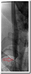
Below is an MRI image of my cervical spine. This is the problem we are trying to solve (or at least halt the further progression of). Arrows indicate MS lesions.

The Bottom Line
I had abnormalities in five out of five veins examined- all are blood vessels that drain my central nervous system. Therefore, it can be said that I had significant CCSVI. The three larger veins were all treated with angioplasty. The two smaller vertebral veins were not treated. One day I hope that we can attempt to treat at least one of these vertebral veins.
Only time will tell if Dr. Sclafani’s efforts were enough to slow or even stop the progression of my MS. I can only hope.
I hope the treatment keeps good news coming.
Judy
Very interesting article, we all hope for the best with you.
Thank you for caring about everyone by posting your journey with MS and now CCSVI. I wish you all the very best and look forward to more good news as it happens for you.
Many thanks for your detailed report. I follow your ride with great excitement hoping to slam the brakes on the progression.
I'm sure that I can speak for everyone else that follows your blog, We are all hoping along with you Mitch. Thanks for the informative post.
Charlie
Many thanks for your detailed report but a follow of your ride definetavely wil be the hope to slam the brakes on the progression of MS. Why is it so difficult to understand this concept?
Mitch this is pretty cool stuff. As always, hoping for the best for you and everyone else that has to put up with the BS of MS. 🙂 Jen
Mitch, very informative stuff. That Mr scalfani did not try to treat those lower veins, Is it due to what is possible to treate at the moment?
Judy, Andy, Bev, ljh, Charlie, Eileen, Jen, thanks for stopping by and leaving such encouraging comments.
Anonymous, yes, Dr. Sclafani didn't balloon the vertebral veins because he's never done that, and does not know of anyone who has.
Thanks for sharing your treatment details (and giving Dr. S the go-ahead to do the same). Really enjoy your blog, and wish you all the best for health and healing.
prairiegirl
Great report with excellent visuals. You will help a lot of people understand what CCSVI is all about. It's really quite remarkable what got done and a day later it feels like nothing.
I've got everything crossed that this halts your progression and that all the veins stay open for years to come!
Me too, Mitch… wishing you the best always.
Lynne
prairiegirl, thanks for the well wishes.
weeble, glad you like the report. That is high praise coming from a former xray technician.
Lynne, thanks for stopping by.
Mitch, I am praying that this all helps. I have many friends that I am sharing your information with. I am PortSports Dave's mom, and he let me know about your "ride". I am enjoying it too.
Riley,
Kim told me that she had shared my website with your son. I'm glad you stopped by to check it out and leave a comment.
We're responsible for sharing information about CCSVI with our MS community, because it's not getting the media attention, or the attention from our physicians, that it should.
Hey Mitch I am glad everything turned out good for you I have read all of your posts and was just wondering did your insurance cover this or did you have to pay for it yourself and if so what does this cost to have done.
Hope everything is fine and good luck But I have to ask the same thing Whats the cost of this
Anonymous and John,
Can't talk $'s in this comments section, but email me by pressing the "click here to email me" button on the right hand side of the blog, near the top.
Mitch,
I notice on the Left Internal Jugular vein images that you posted , the stenosis is present on the post balloon dilation image (just beside where the collar joins the breastbone). Did you post the incorrect image?
Chiefumtaga,
That is the correct image, and according to my physician it shows improved blood flow, not post procedure stenosis. Kind of like trying to determine the sex of your baby by looking at an ultrasound yourself…